Arm Blood Vessels Labeled : Arteries Nerves And Muscles Of Upper Limb Anterior View Muscles Of Forearm Deep Layer Anterior View / The ruptured or broken blood vessels on arm may be more of a cosmetic issue than a medical condition.
byAdmin•
0
Arm Blood Vessels Labeled : Arteries Nerves And Muscles Of Upper Limb Anterior View Muscles Of Forearm Deep Layer Anterior View / The ruptured or broken blood vessels on arm may be more of a cosmetic issue than a medical condition.. The forearm region is thus supplied by two major vessels, the radial artery and ulnar artery. In terms of arterial supply, the upper limb has 5 main vessels, the: The anterior cubital fossa, visible with the integumentary layer faded in complete anatomy. Know the causes, types, symptoms and home remedies for broken blood vessels on arm. It has only one bone called the humerus, and an intricate network of muscles, vessels and nerves distributed around it.
Blood is supplied to parts within the neck, head and brain through branches of the subclavian and common carotid arteries. The profunda brachii terminates by contributing to an anastomotic network around the elbow joint. The first part is an extension of the subclavian artery. The vessels of the arms are part of the circulatory system, which provides nutrients to the tissues. Ecg and pulse (including effect of cold temperature and raising arm (gravity) on pulse amplitude) taking apical pulse.
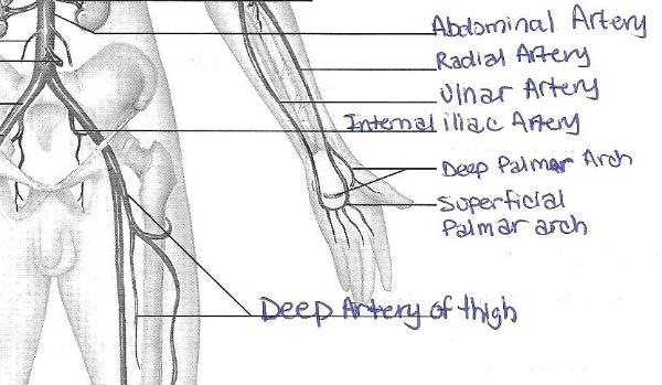
Wire Models from classroom.sdmesa.edu Which vessel is missing from the following statement tracing blood that drains from the large intestine, we find that blood drains from the distal colon is collected in the inferior mesenteric vein, merges with the splenic vein then directed to the hepatic portal vein, the liver sinusoid and the inferior vena cava Slide of artery, vein, and capillaries. The arteries and veins are a vast network of blood vessels responsible for carrying blood throughout the entire body. Arteries (in red) are the blood vessels that deliver blood to the body. It allows blood to flow into the arm, but prevents all of it from. In the forearm, it travels deep under muscle along the small finger side of the forearm. Around the t4 vertebra the aortic arch transitions into the thoracic aorta. The vessels which supply the wrist and hand are the ulnar and radial arteries.
The following events may cause thoracic outlet syndrome, especially in people with the above bone or muscle abnormalities in the neck:
Download 73 arm vessels stock illustrations, vectors & clipart for free or amazingly low rates! The axillary artery is a blood vessel that provides the axilla, the lateral portion of the thorax, and the upper limb with oxygenated blood. Anatomy, human arm with muscles, nerves, blood vessels and bones / anatomie, menschlicher arm mit muskeln, nerven, blutgefäßen und knochen, historisch, historical, digital improved reproduction of an original from the 19th century / digitale reproduktion einer originalvorlage aus dem 19. Shoulder and arm label the blood vessels of the right arm. It extends from the shoulder joint to the fingers and contains 30 bones. This process of blood flow within your body is called circulation. The brachial artery proper descends down the arm. The blood supply to the deep flexor and extensor muscles of the forearm is supplied via the ulnar artery that divides into anterior and posterior interosseous arteries, on the dorsal side of the wrist, there is another merger of these arteries that of which forms the carpal arch; The cephalic, median antebrachial, and basilic veins carry blood through the arms until they join the deep veins at the brachial vein. The delineation between the subclavian artery and the axillary artery is the lateral most border. A tourniquet is an elastic strap tied around the arm proximal to the venipuncture site. The ruptured or broken blood vessels on arm may be more of a cosmetic issue than a medical condition. We'll go over the bones, joints, muscles, nerves, and blood vessels that make up the human arm.
Three blood vessels branch from the aortic arch: The nerves of the arm are supplied by one of the two major nerve plexus of the human body, the brachial plexus. The aortic arch is the beginning of the aorta, where it exits the left ventricle. The common cartoid artery extends from the brachiocephalic artery. The cephalic, median antebrachial, and basilic veins carry blood through the arms until they join the deep veins at the brachial vein.
Arm Veins Diagram Quizlet from o.quizlet.com A tourniquet is an elastic strap tied around the arm proximal to the venipuncture site. The delineation between the subclavian artery and the axillary artery is the lateral most border. Shoulder and arm label the blood vessels of the right arm. The anterior cubital fossa, visible with the integumentary layer faded in complete anatomy. Slide of artery, vein, and capillaries. Which vessel is missing from the following statement tracing blood that drains from the large intestine, we find that blood drains from the distal colon is collected in the inferior mesenteric vein, merges with the splenic vein then directed to the hepatic portal vein, the liver sinusoid and the inferior vena cava It consists of three sections, the upper arm, forearm, and hand. The brachiocephalic artery, the left subclavian artery, and the left common carotid artery.
Blood is supplied to parts within the neck, head and brain through branches of the subclavian and common carotid arteries.
The axillary artery supplies blood to the lateral aspect of the thorax, axilla (armpit), and upper limb. The brachial artery proper descends down the arm. Labeled arm showing the antecubital veins / normal blood pressure is 120/80. The forearm region is literally full of muscles, with twenty of them laying within two compartments, all requiring a rich blood supply. The iliac, femoral, popliteal and tibial (calf) veins are the deep veins in the legs. Any of these abnormal formations can compress blood vessels or nerves. Arm and hand symptoms that persist long after a whiplash injury may be a sign of thoracic outlet syndrome. Blood is supplied to parts within the neck, head and brain through branches of the subclavian and common carotid arteries. Arm blood vessels labeled : It consists of three sections, the upper arm, forearm, and hand. The ulnar artery comes from the brachial artery and travels across the front of the elbow. The brachiocephalic artery, the left subclavian artery, and the left common carotid artery. In the forearm, it travels deep under muscle along the small finger side of the forearm.
They face much lower blood pressures than. The arteries and veins are a vast network of blood vessels responsible for carrying blood throughout the entire body. The forearm region is thus supplied by two major vessels, the radial artery and ulnar artery. The brachiocephalic artery, the left subclavian artery, and the left common carotid artery. Vessels on torso, thorax, and pelvis models.
Exercise 32 Anatomy Of Blood Vessels Flashcards Easy Notecards from www.easynotecards.com Katy wallis at state college of florida The iliac, femoral, popliteal and tibial (calf) veins are the deep veins in the legs. The cephalic, median antebrachial, and basilic veins carry blood through the arms until they join the deep veins at the brachial vein. The nerves of the arm are supplied by one of the two major nerve plexus of the human body, the brachial plexus. Blood is supplied to parts within the neck, head and brain through branches of the subclavian and common carotid arteries. The axillary artery is a blood vessel that provides the axilla, the lateral portion of the thorax, and the upper limb with oxygenated blood. The delineation between the subclavian artery and the axillary artery is the lateral most border. The arm muscles are divided into two compartments separated by the humerus and the medial and lateral intermuscular.
The profunda brachii terminates by contributing to an anastomotic network around the elbow joint.
They face much lower blood pressures than. It extends on each side of the neck and divides at the level of the larynx into two branches: Arm and hand symptoms that persist long after a whiplash injury may be a sign of thoracic outlet syndrome. The brachiocephalic artery, the left subclavian artery, and the left common carotid artery. This artery comes close to the skin surface. Ecg and pulse (including effect of cold temperature and raising arm (gravity) on pulse amplitude) taking apical pulse. Labeled arm showing the antecubital veins / normal blood pressure is 120/80. However, for some, broken blood vessels can be a cause of concern and may indicate an underlying medical condition. It consists of three sections, the upper arm, forearm, and hand. The blood supply to the deep flexor and extensor muscles of the forearm is supplied via the ulnar artery that divides into anterior and posterior interosseous arteries, on the dorsal side of the wrist, there is another merger of these arteries that of which forms the carpal arch; The arm muscles are divided into two compartments separated by the humerus and the medial and lateral intermuscular. The radial artery travels across the front of the elbow, deep under muscle until it comes to the wrist. Three blood vessels branch from the aortic arch:
They face much lower blood pressures than blood vessels labeled. The common cartoid artery extends from the brachiocephalic artery.